- Document History
- Subscribe to RSS Feed
- Mark as New
- Mark as Read
- Bookmark
- Subscribe
- Printer Friendly Page
- Report to a Moderator
- Subscribe to RSS Feed
- Mark as New
- Mark as Read
- Bookmark
- Subscribe
- Printer Friendly Page
- Report to a Moderator
*Visit the official "Submit Entry" page to enter your NI LabVIEW student design project in the competition*
*Please add your country to the title of your project, example: Amazing Robot Project, Poland.
Contact Information
University: Korea University, Department of Electronics and Information - Medical Imaging System & Science Laboratory
Team Member(s): Chang-Hyun Oh, Jeung-Hoon Seo, Sang-Doc Han, Jeun-Seon Jeong, Jae-Hoon Kim, Sang-Woon Han, Hyun-Ku Lee, Jun-Hyuk Kim, Min-Gyu Hyeon, Ewi-Don Jeong, Ki-Won Kim, Bum-Seok Kim, Sung-Yeon Cho, Min-Woo Choi, Jeong-Wook Choi, Sung-Min Lee, Sun-Hong Kim, Young-Gyu Lee, Ki-Jeong Bang, Hye-Jin Seong, Do-Hyun Kim, Sun-A Lee, Hyeong-Joo Jo, June-Soo Park, Jae-Mu Kim, Jea-won Huh
Faculty Advisors: Chang-Hyun Oh
Email Address: ohch@korea.ac.kr,jhseo-mri@korea.ac.kr, koreatap@korea.ac.kr, kslove162@korea.ac.kr, allzz@korea.ac.kr, sangwoon088@korea.ac.kr, fufuru09@korea.ac.kr,sura6404@korea.ac.krfufuru09@korea.ac.kr, sura6404@korea.ac.kr,fufuru09@korea.ac.krfufuru09@korea.ac.kr, sura6404@korea.ac.kr,, sura6404@korea.ac.kr, rhrlmando@korea.ac.kr, jewidon@korea.ac.kr, supershutank@korea.ac.kr, jewonjewon@korea.ac.kr, kbs7452@korea.ac.kr,syluxury@korea.ac.krkbs7452@korea.ac.kr,syluxury@korea.ac.krkbs7452@korea.ac.krkbs7452@korea.ac.kr,syluxury@korea.ac.kr,syluxury@korea.ac.kr, kingarioo@korea.ac.kr, esuerid@korea.ac.kr, sinsomy@korea.ac.kr, salong@korea.ac.kr, notion114@korea.ac.kr, rlwjd1218@korea.ac.kr, domeuu@korea.ac.kr, dohyun704@korea.ac.kr, lsa1297@korea.ac.kr, shutupcj@korea.ac.kr, pjsu21@korea.ac.kr, myper@korea.ac.kr
Country: Korea
Project Information
Title: Image Reconstruction and processing for portable MRI
Description: As a part of a small portable MRI system with a labview-oriented console and spectrometer, we have developed a package to reconstruct MR images and to improve images.
Products:
(1) Labview Software with 2010
(2) RIO (NI) SBRIO -9602
(3) RF unit (Radio Processor and NI USRP-2920)
The Challenge: (1) Spectrometer for MRI : ( i) with low frequency signal outputs (three, DC to 5 kHz signals) (ii) RF outputs (approximately 100 kHz to 100 MHz arbitrary waveform generator) (iii) RF inputs with a frequency same as (ii) to be demodulated and used as a raw data to reconstruct MR images. (iv) Post processing software for MR images
The Solution:
<insert explanation of how your project works>
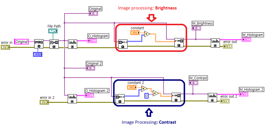
(1) Hardware driving software (RF and gradient signal)
(2) Hardware acquisition software (RF)
(3) 3-D reconstruction for for MRI (3-D Fourier transform program with various options)
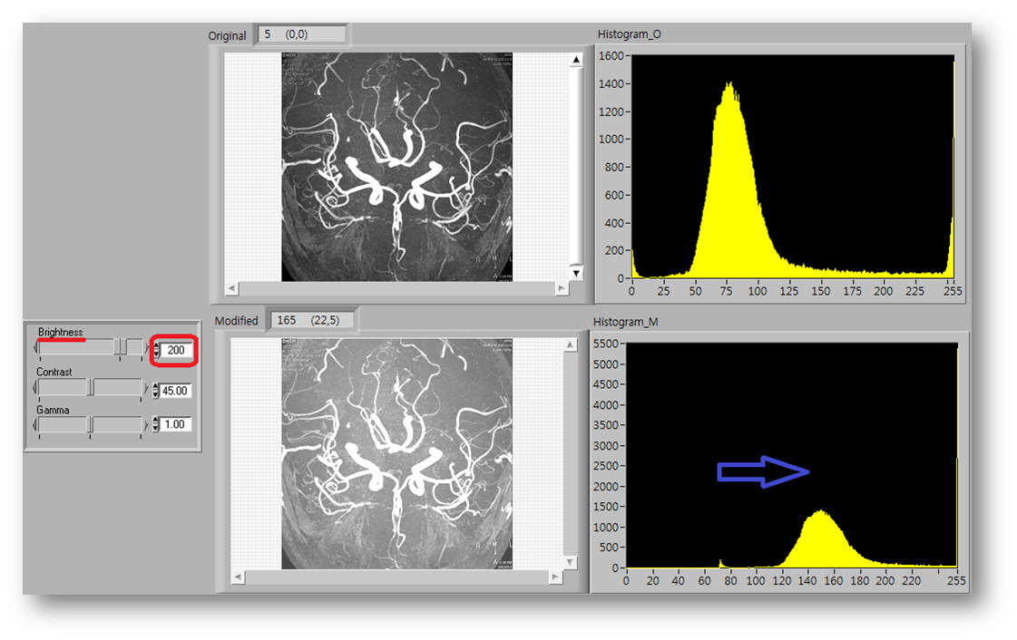
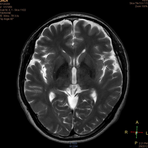
(4) Image processing software to enhance and understand MR images.
<insert explanation of the benefits gained using LabVIEW and NI tools>
(1) Hardware Accessibility
(2) Easy to understand complex digital image processing and Reconstruction Algorithm
(3) Good as a starting project to develop systems with more options such as medical imaging systems with more complexity. (later in a company etc.)
<insert image(s) of project with captions>
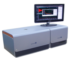
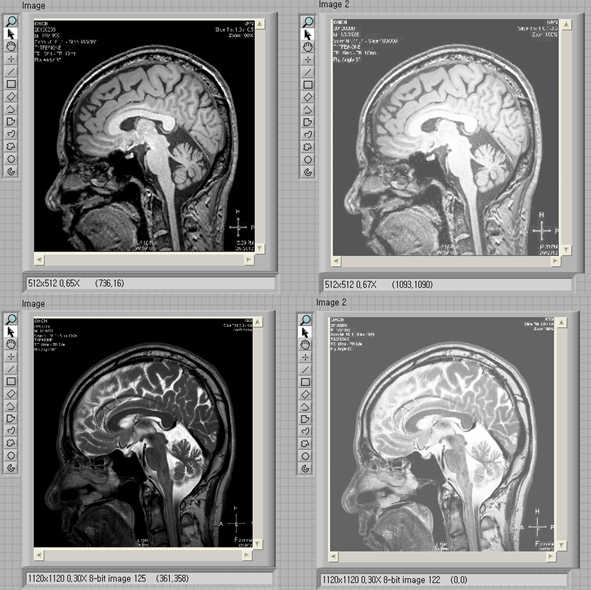
Target MRI system
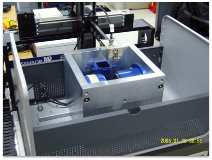
Inside view of the system showing the magnet, gradient/shimming coil and RF coil to acquire MR signals
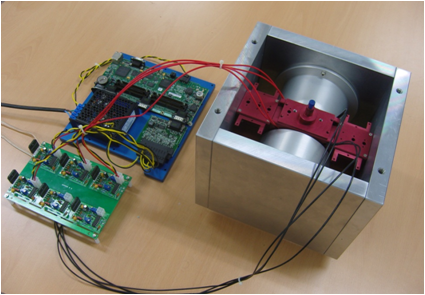
Picture of the magnet and shim/gradient coils
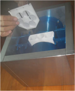
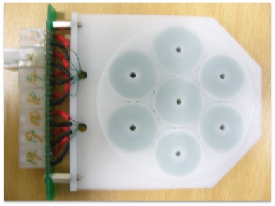
Cassette-Type shim-gradient coil
<reminder: attach VI code (optional)>
(1) Explanation and Source Code for 3-D MRI Reconstruction:
Explanation: 3-D MR image reconstruction is done using a 3-D discrete Fourier tranform with various optional parameters such as zero-appending options, filtering options, image direction change, and selection options with various ROI’s.
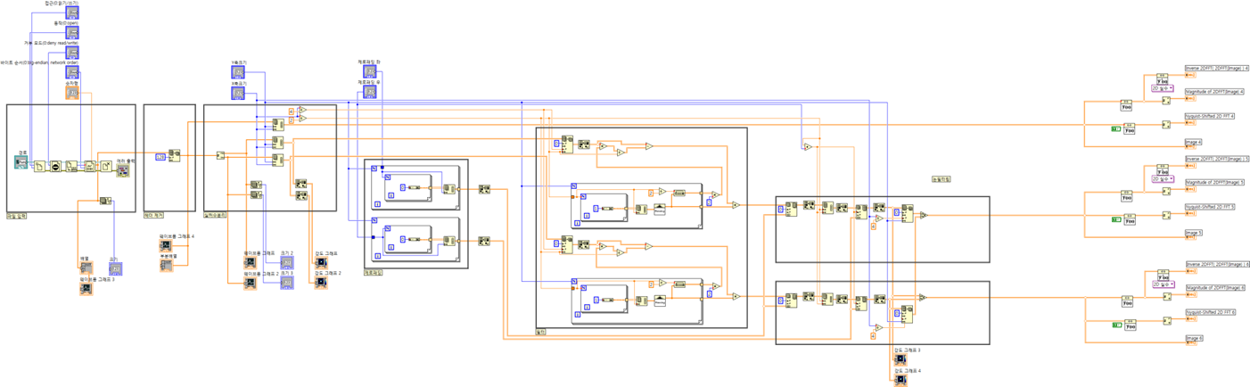
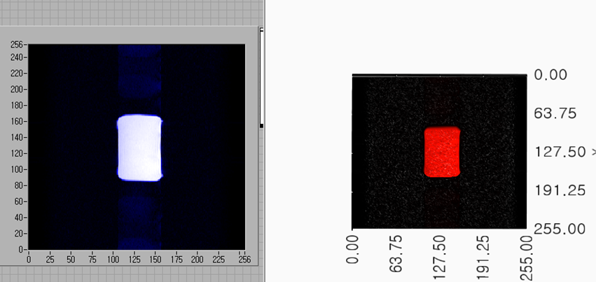
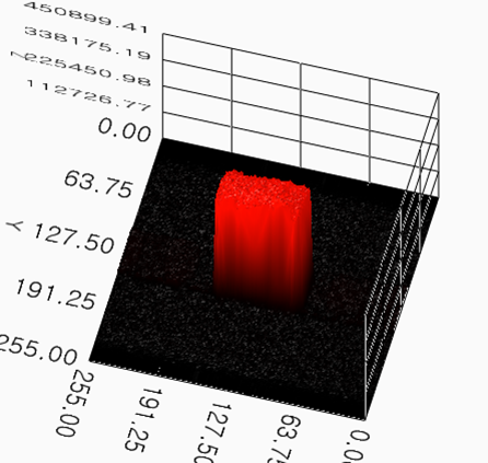
VI Code 2-D MRI Reconstruction Image 3-D MRI Reconstruction Image
(2) List of Processing Methods for MR images and some of source code
List:
a. Image Negatives
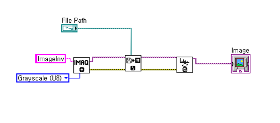
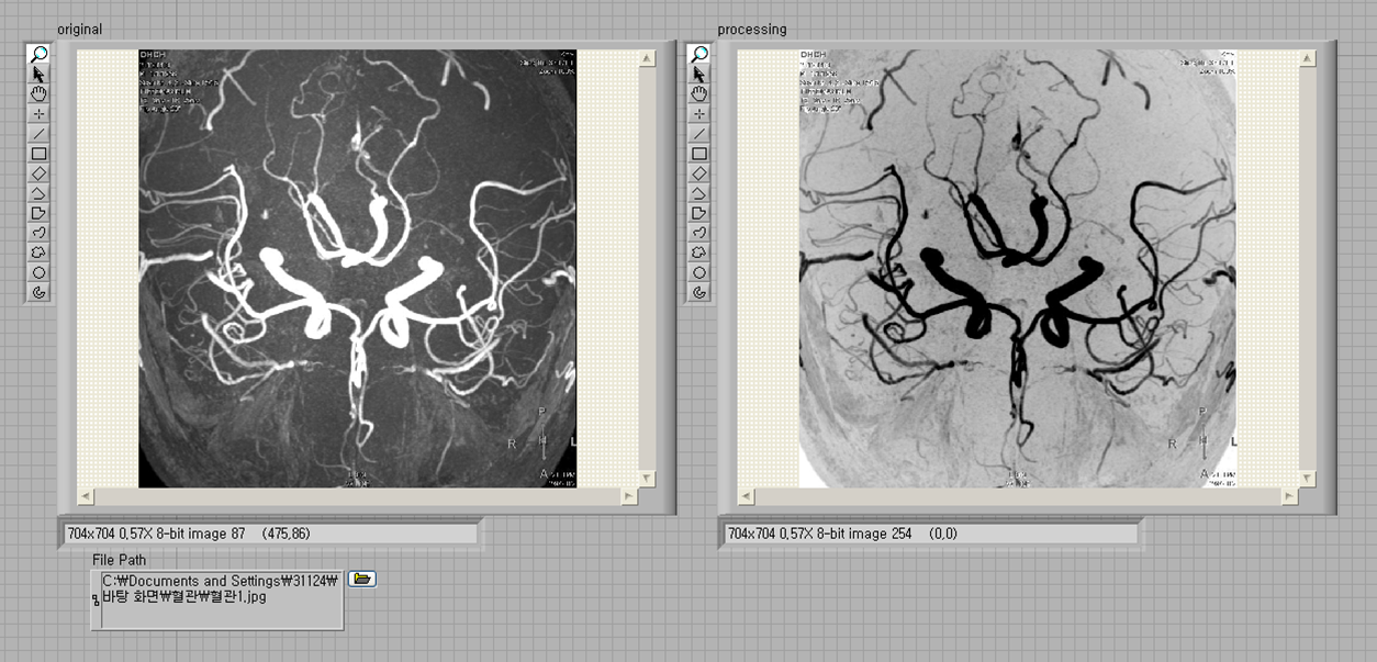
VI Code original Image porcessed Image
b. Gamma and Log compensation
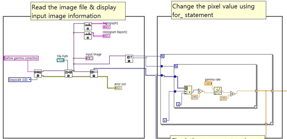
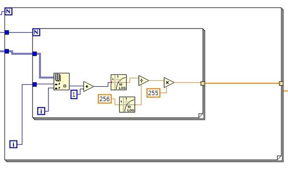
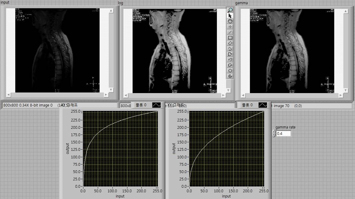
VI Code original Image porcessed Image
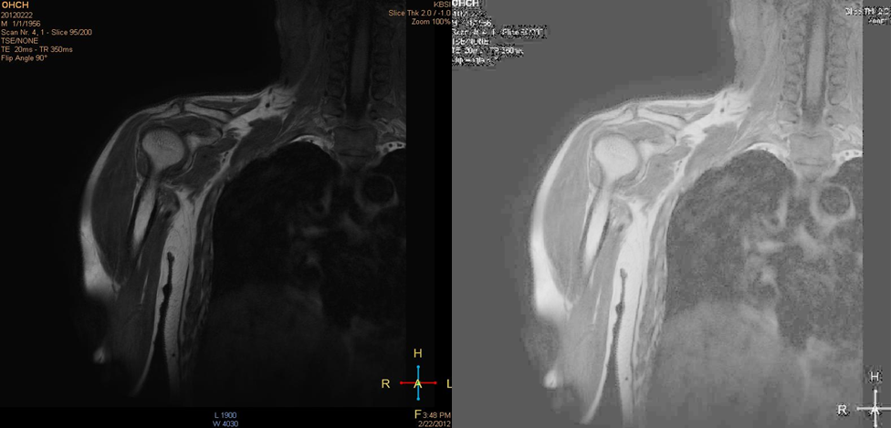
c. Contrast Stretching
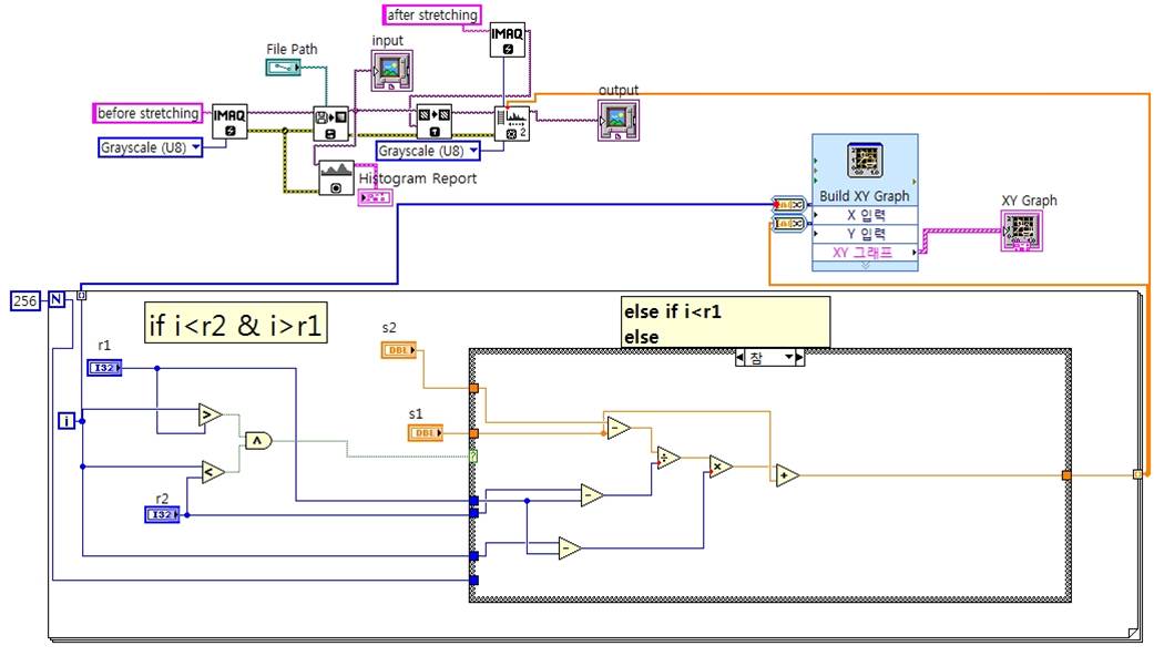
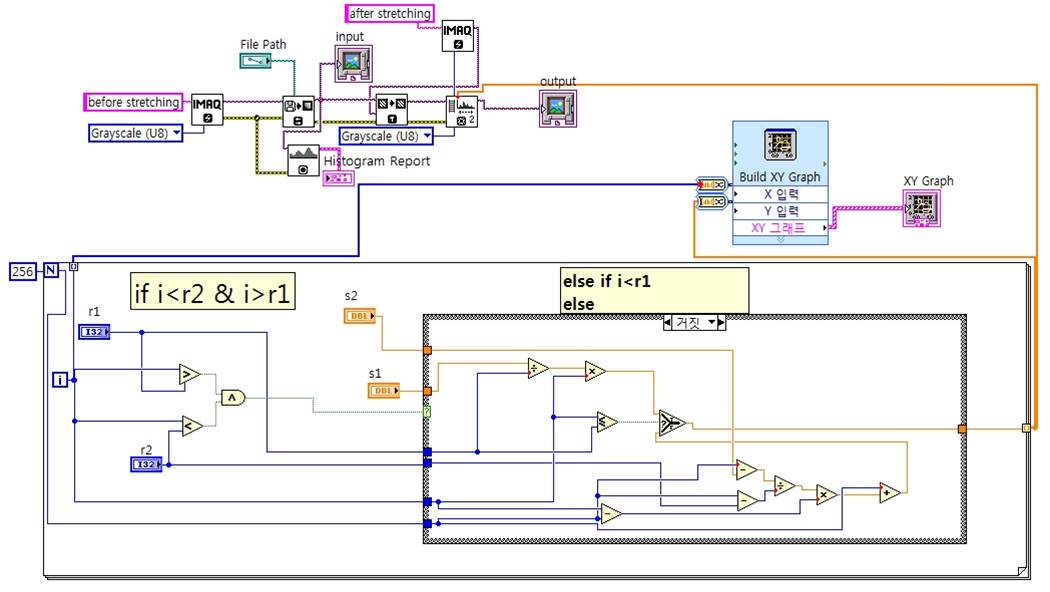
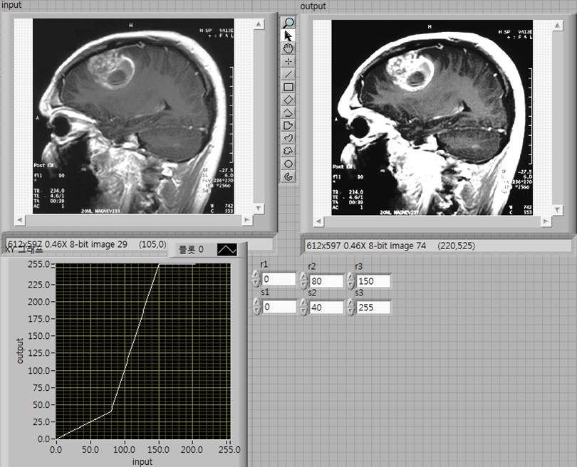
VI Code original Image porcessed Image
d. Making Basic Image Types
VI Code top original Image bottom porcessed Image
e. Histogram Equalization
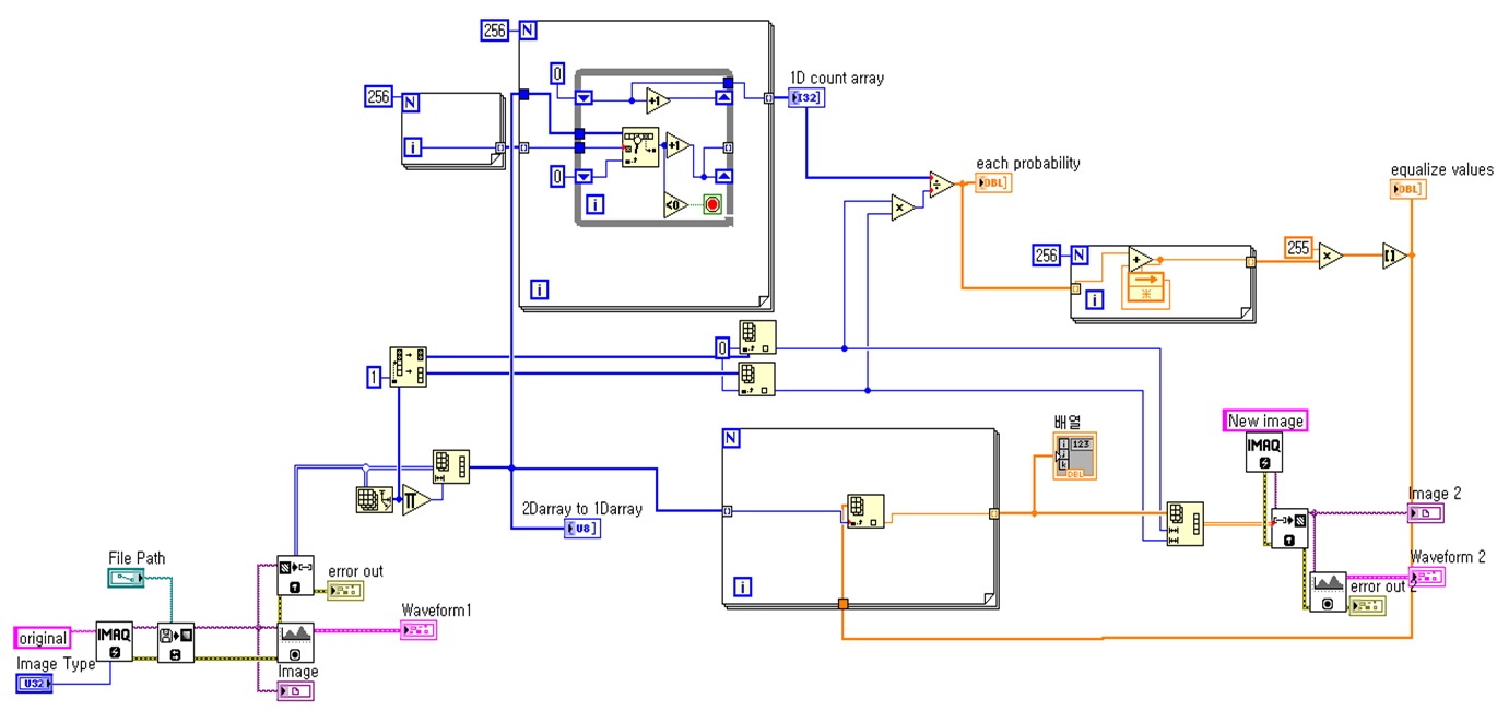
VI Code original Image porcessed Image
f. Laplacian
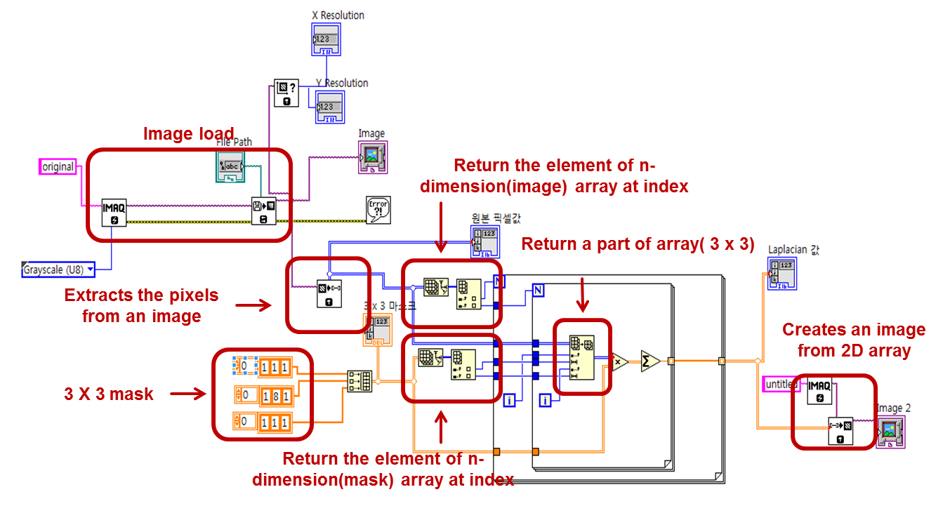
VI Code original Image porcessed Image
g. Unsharp Masking
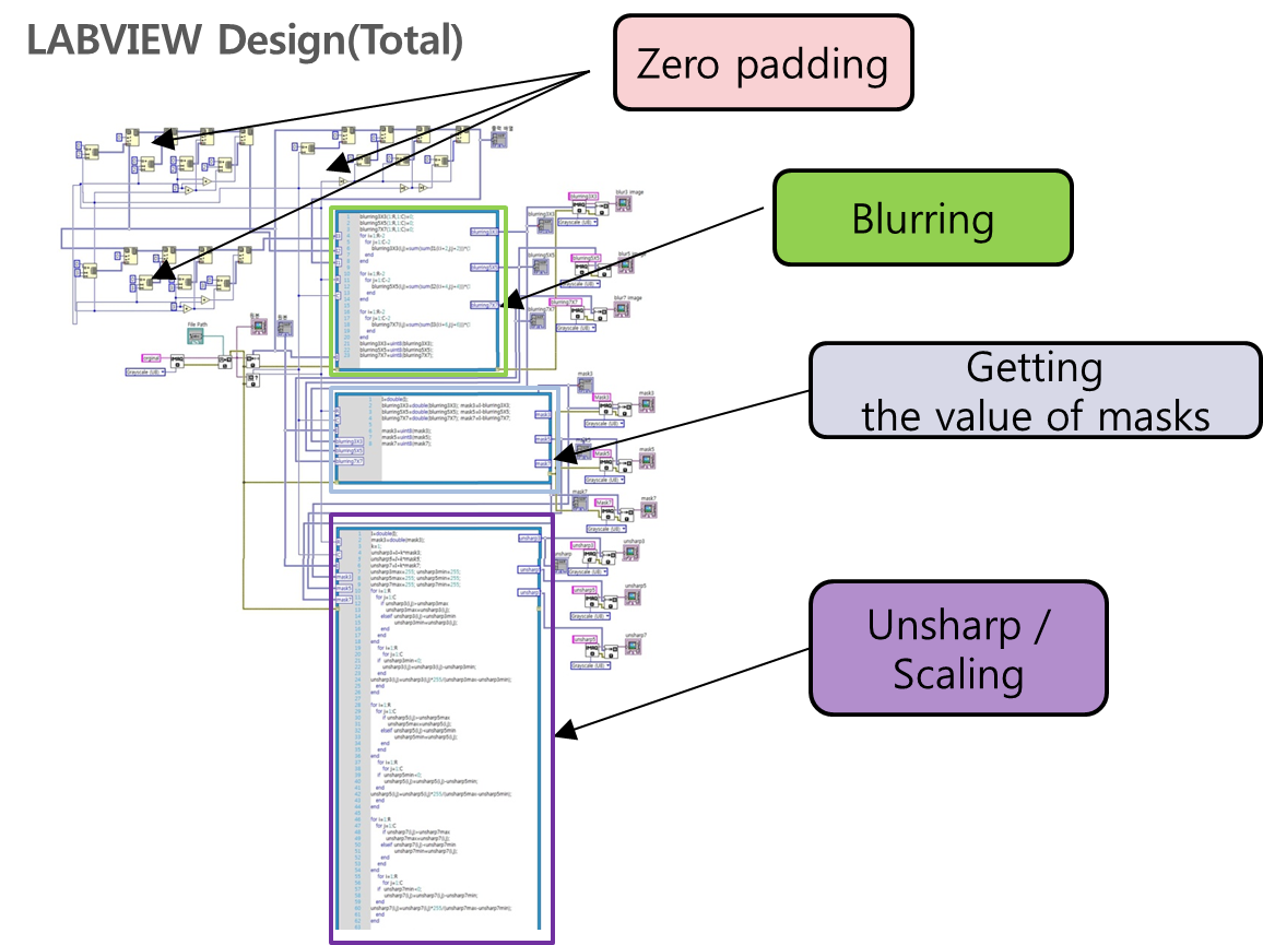
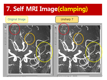
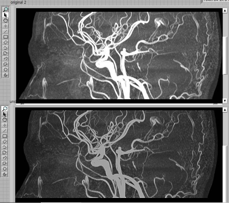
VI Code original Image porcessed Image
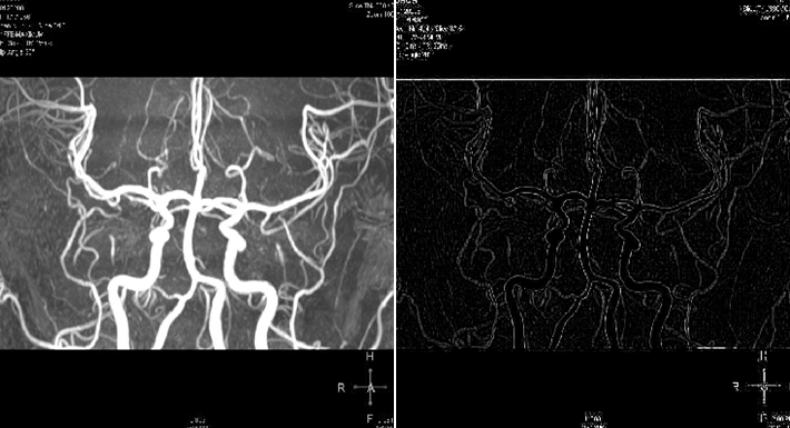
h. Gradient
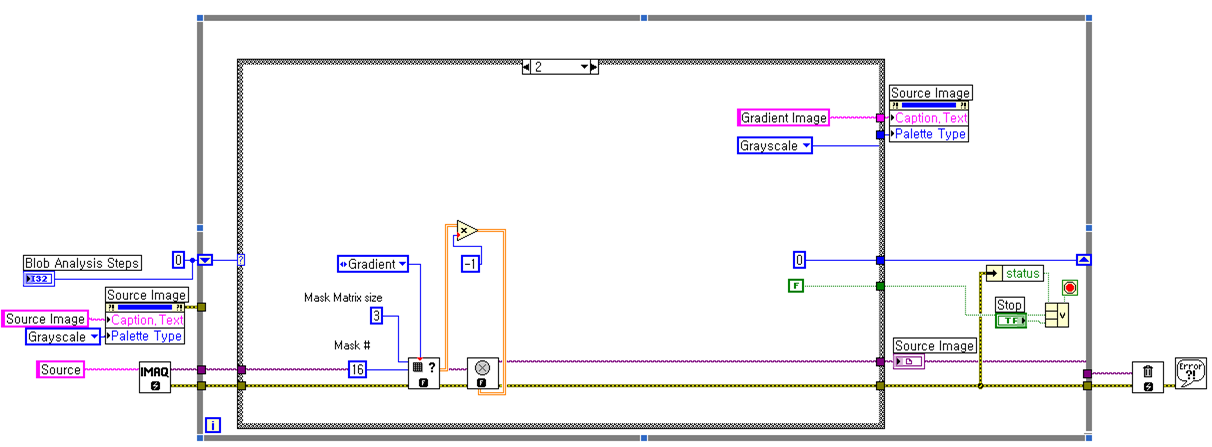
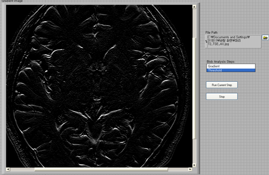
VI Code original Image porcessed Image
i. Ideal Low Pass Filter
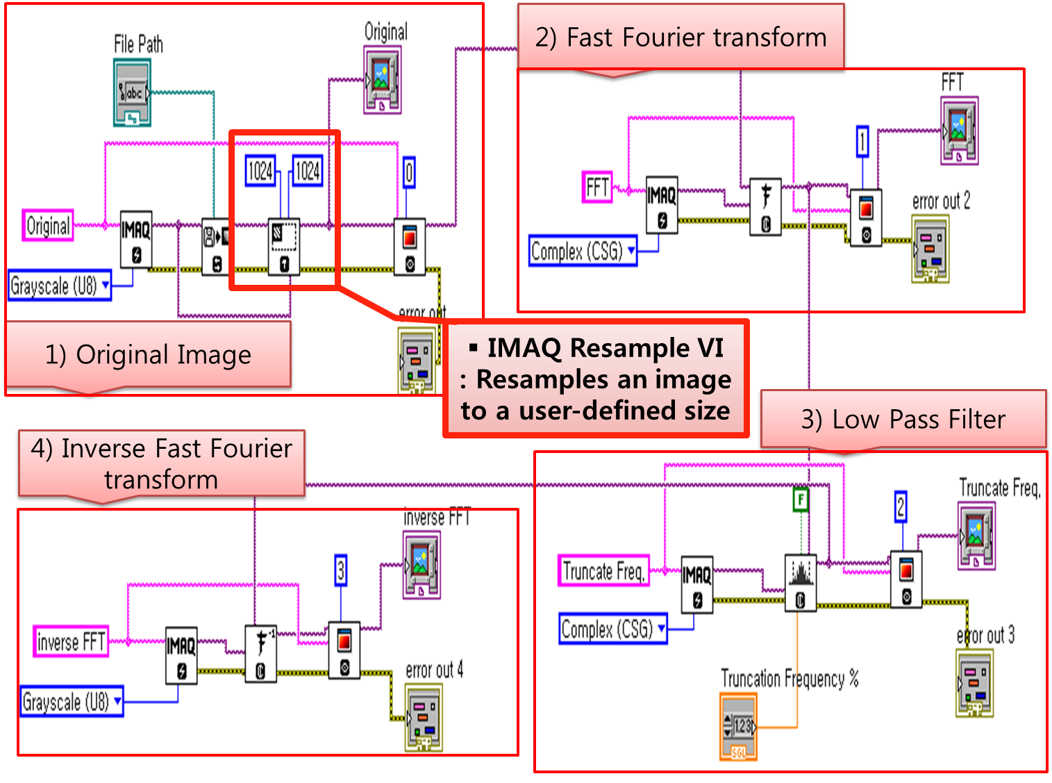
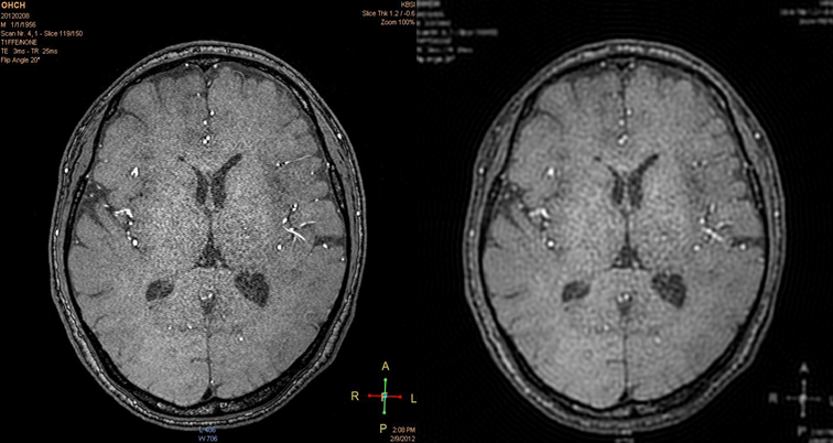
VI Code original Image porcessed Image
Nominate Your Professor: (optional)
<insert nomination. Does your professor use LabVIEW or other National Instruments technology to make learning difficult concepts engaging, interesting, and fun? If so, nominate him/her as an outstanding educator by telling us who they are, what they teach, and how they make learning a better experience for you.>
- Mark as Read
- Mark as New
- Bookmark
- Permalink
- Report to a Moderator
Great work I had ever see.
