- Document History
- Subscribe to RSS Feed
- Mark as New
- Mark as Read
- Bookmark
- Subscribe
- Printer Friendly Page
- Report to a Moderator
- Subscribe to RSS Feed
- Mark as New
- Mark as Read
- Bookmark
- Subscribe
- Printer Friendly Page
- Report to a Moderator
Diffraction Imaging of Micro-particles and Bacteria using LabVIEW
Contact Information
University: University of South Florida
Team Member (with year of graduation): Karthik raj Konnaiyan (2015)
Project Supervisor: Dr.Anna Pyayt
Email Address: karthikrajk@mail.usf.edu
Project Information
Title: Diffraction Imaging of Micro-particles and Bacteria using LabVIEW
Description: Shape recognition of micro-scale objects using NI LabVIEW processing of laser diffraction patterns.
Products: LabVIEW 2013, NI Vision Acquisition, NI Vision Assistant, Logitech Webcam
The Challenge:
The U.S. Centers for Disease Control and Prevention (CDC) estimated that 2 million illness and 23,000 deaths are associated with bacterial infections. There is a need for fast detection and identification of bacteria allowing to minimize negative consequences of the infection. There are number of methods for bacterial detection, which include gram staining or culturing followed by further analysis that is usually very time consuming. We propose to use image processing of real time videos demonstrating laser diffraction on potentially contaminated samples for instant, low cost, label-free bacteria detection.
The Solution:
A green laser was illuminating low concentration bacteria sample deposited on a glass slide. The images of the diffracted pattern were captured by a web camera. Samples containing micro beads, rod and spiral shaped bacteria were used in the study. The diffracted images of the sample were then transferred into the computer by NI Vision Acquisition and analyzed by NI’s Vision Assistant to check the presence of bacteria or micro bead and differentiate them based on their morphologies. This process was done by extracting luminance plane (grayscale) from the color image. A particle classifier tool was used to examine the bacteria structures and differentiate them by comparing the structures with the pattern template that has been stored in the database. A program in the LabVIEW was created to store the particle classifier information and create a report describing content of a specific sample.
National Instruments Vision Assistant software was used for processing of the images. The specific tools included Color plane extractor, lookup table and particle classifier. The image obtained by the web camera was streamed into the computer. Once the image was read into the software the luminance plane (grayscale image) was extracted from the color image. This preprocessed image was analyzed using particle classifier detecting bacterial presence as well as identifying the its structure. This was done by comparing with bacterial templates of like rod-shaped, spiral-shaped and spherical structures that were stored in the database. The accuracy was improved by increasing the number of erosion which corresponded to removal of background noise.
Figure 1: Experimental setup - Lateral view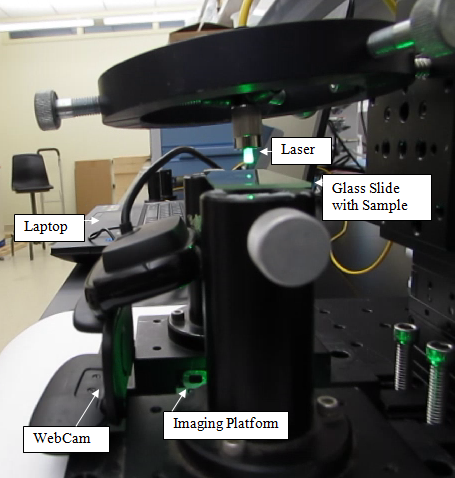
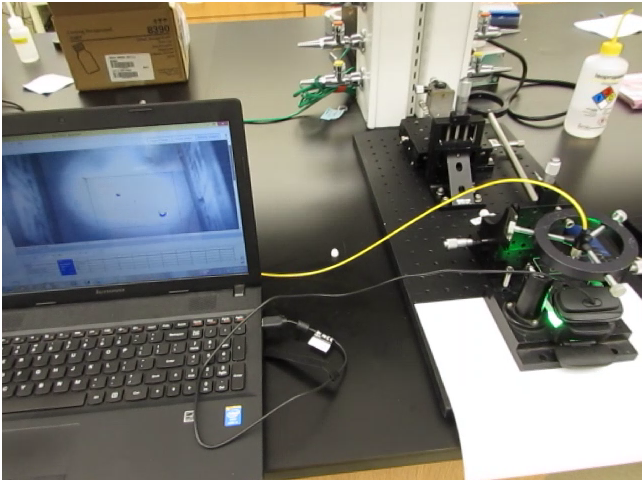
Figure 2: Experimental setup- front view

Figure 3:Preprocessed image by LabVIEW to vizualize the bacteria-NI Vision Assistant
Figure 4: Detection of Spirillum-NI Vision Assistant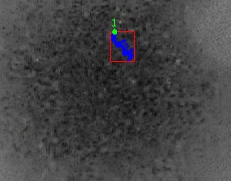
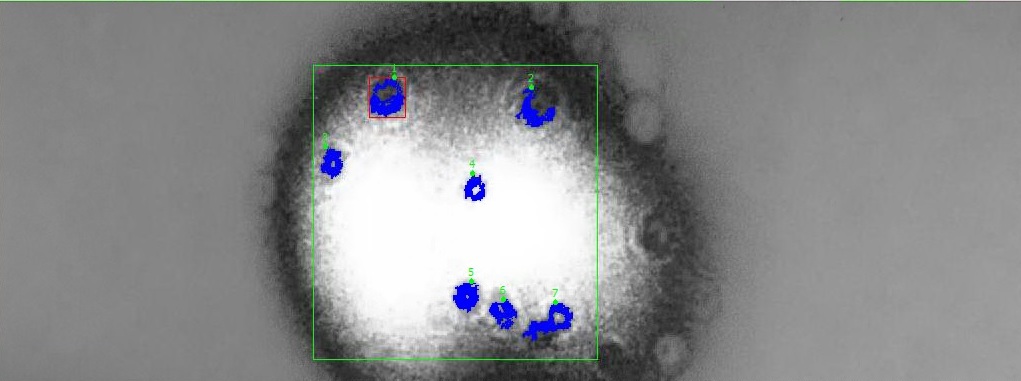
Figure 5: Detection of Microbeads-NI Vision Assistant
This project is currently in alpha level which requires more samples to be trained for a large database.
This project took 5 months to complete
Attachment:
- Experimental setup.jpeg
- Experimental setup- front view.jpeg
- Experimental setup - Lateral view.jpeg
- Preprocessed image by LabVIEW to visualize the bacteria-NI Vision Assistant.jpeg
- Detection of Spirillum-NI Vision Assistant.jpeg
- Detection of Microbeads-NI Vision Assistant.jpeg
- Front panel - Diffraction Imaging of Micro-particles and Bacteria.jpeg
- Block diagram of Image processing module.jpeg
- Poster.pdf
- Project Video
I like to nominate my Professor Dr.Anna Pyayt who motivated me to design this project. Apart from this she is an outstanding educator who makes the class interesting by giving different kind of interesting projects which gave opportunities to many for building their career. i am proud to be one among the group.
- Mark as Read
- Mark as New
- Bookmark
- Permalink
- Report to a Moderator
Great work.
