- Document History
- Subscribe to RSS Feed
- Mark as New
- Mark as Read
- Bookmark
- Subscribe
- Printer Friendly Page
- Report to a Moderator
- Subscribe to RSS Feed
- Mark as New
- Mark as Read
- Bookmark
- Subscribe
- Printer Friendly Page
- Report to a Moderator
Contact Information
Country: Switzerland
Year Submitted: 2018
University: Swiss Federal Institute of Technology (ETH) Zurich
List of Team Members: David Mauderli, Oliver Steffen, Yves Haag, Simon Rogler, Georg Brunner, Simona Santamaria
Faculty Advisers: Dr. Prof. Mirko Meboldt, Jan Zimmermann, Bente Thamsen
Main Contact Email Address: ssimona@ethz.ch
Project Information
Description: This project developed a simulator for surgeons to train minimally invasive heart operations. With realistic haptic feedback and simulated imaging technologies from the operating theatre, the surgeons will get the best training to do a transseptal puncture procedure.
Products:
NI CompactRIO-9034 with modules NI 9472, NI 9421, NI 9401, working with LabVIEW 2017 and the NI Vision Acquisition Software.
The Challenge
In the last few years simulation based training methods have become increasingly important concerning education of medical personnel. This is mainly due to the fact that training on a simulator is far less dangerous than training on living patients. In the field of cardiovascular interventions there is still plenty of room for better high-fidelity simulation tools. This project aims to close this gap by developing a simulator for the transseptal puncture procedure, focusing on a realistic haptic feedback.
The Solution
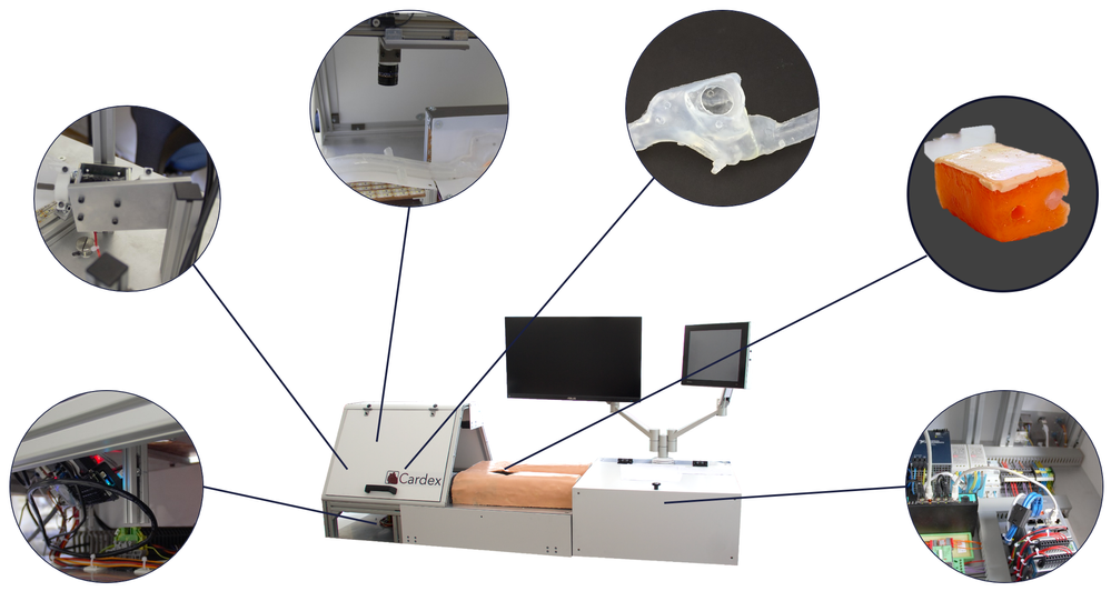
We focused on developing a product that is easy and intuitive to use. But hidden from the user’s eyes there are many parts and systems that make all this possible. In the following the most important features are explained while also going through the main steps of a transseptal puncture procedure.
Systems overview
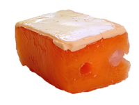
The first step is to achieve access to the veins at the groin. After identifying the correct spot, the hollow needle is inserted. For our Simulator, we developed and manufactured a special PVA pad. Its two layers mimic the skin and the tissue below.
Insertion Pad
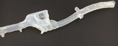
Continuing the operation, the guidewire is pushed through the needle and advanced into the venous system. In our Simulator, the venous system and heart are replaced by a silicone model. We took Ct-Scans of a real patient’s heart and processed it to be used in our simulator. Because after molding the silicone still has a very high friction, it is specially treated, so the guidewire does not get caught up.
Our phantom
In the next step, the hollow needle is removed. In its place, the catheter is pushed over the guidewire. As the surgeon doesn’t have direct vision anymore he activates x-ray with the foot pedal to confirm the position. Real X-Ray as used in the operation theatre emits radiation, which is harmful for both, the surgeon and the patient. We are proud to say that we have developed a solution using carefully crafted LED panels and cameras, that can sufficiently mimic this imaging technique. All without ever exposing the learning surgeon to harmful radiation.
The left picture shows the real x-ray, the right one is our x-ray simulation.
Once at the heart, the guidewire is removed and the transseptal needle is inserted in its place. Following is the most difficult and critical step. The surgeon slowly pulls down the catheter. During this he is constantly checking x-ray for two jumps the catheter makes. One when it falls into the atrium, and the second, when it reaches the soft tissue of the Fossa Ovalis. Using Ultrasound imaging he looks for the so-called tenting, a bending of the Fossa Ovalis.
Puncturing the correct spot is of the utmost importance, because only small deflections could mean the surgeon is puncturing outside the heart or even worse the aorta, which could have severe consequences or even be fatal for the patient. Therefore, we aimed to incorporate this imaging technique as well.
Ultrasound imaging as used in the operation theater only works with fluids. In our simulator, we wanted to avoid using fluids, to reduce the risks of spills and make the user experience more pleasant. Therefore, we developed a scanning system based on a stereo camera setup, that constantly measures the deformation of the Fossa Ovalis and transforms this Information into a classical ultrasound image.
The left picture shows the real ultrasound, the right one is our ultrasound simulation.
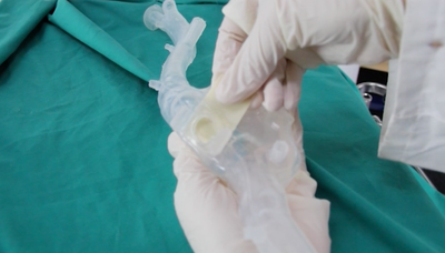
Once the surgeon is at the correct position he advances the trasseptal needle and punctures the membrane. As the Fossa Ovalis is destroyed in this process, we developed a card and slot system, that enables the surgeon to exchange the Fossa Ovalis in mere seconds.
The card and slot system to replace the Fossa Ovalis
By clicking “punctured” on our user interface, the surgeon completes the session and is provided feedback on how well he fared. This way he can objectively track his improvement and build confidence for the real procedure.
The operation is very complicated. In the following video the explained steps are visualised.
It was very important for us to have a realistic simulation of the real x-ray and ultrasound, used in the operating theatre. The CompactRIO whit its real-time operating system and Vision Acquisition Tool enabled us to sufficiently mimic this imaging technologies. The programming language of LabVIEW is very graphical and helped a lot to have an overview of the code. Our simulator includes a monitor and a touch panel for the user, different Network communications and already implemented SubVI’s as well as NI tools were an incredible help to finalise our system. The following graph shows an overview of our mechatronics components.
Mechatronics Overview
Conclusion
We are proud to say that surgeons can start to train on our simulator. We, a group of six mechanical engineering students, managed to build a simulator in nine months.
Our team


